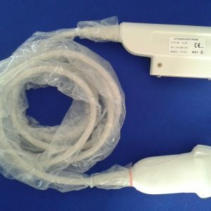
When comparing transvaginal probes and ultrasounds, you might wonder if they are better for your body. This article will discuss the benefits of transvaginal ultrasounds, why they are better, and how they can transmit HPV. Before you get started, make sure your bladder is empty. Before you begin your exam, be sure to follow these important tips:
The debate on whether
transvaginal probes are better than transrectal ultrasound scans has lasted longer than the technology itself. In one study, the researchers used a Philips/ATL 3000 system in Bothell, WA and a GE Logiq 700 system in Milwaukee, WI. All patients underwent TRS after hearing the explanation of the procedure. The vaginal probe was covered with a customary plastic sheath and lubricated with KY gel. The scanning method was similar to that of TVS.
The effectiveness of the new
transvaginal ultrasound procedure depends on a few factors. First, it must be sterile. Using an unsterile
transvaginal probe for ultrasound examinations can cause infection and other complications. Changing the probe between each procedure can make the procedure more dangerous for the patient. A new study by the American College of Obstetrics and Gynecology indicates that transvaginal ultrasound is safe when done by a certified physician.
Another important factor to consider is how to clean the transvaginal
ultrasound probe. The recommended cleaning procedures vary depending on the classification of the ultrasound probe. Cleaning can range from simple wiping to sterilization. Semi-critical devices are at a higher risk of transmission of infections because they come into contact with mucous membranes and non-intact skin. For these reasons, high-level disinfection is recommended.
They provide better resolution
While transvaginal ultrasound is well-known for gynecologic diagnosis, there is now evidence that this technology can be used in other organ systems. With the introduction of high-resolution transducers, transvaginal US provides excellent resolution and can be placed close to the uterus and ovaries for improved diagnosis. High-resolution transducers are especially helpful in examining the vascular supply of the pelvic organs, including the uterus and ovaries. Transvaginal ultrasound can also provide detailed images of the foetal anatomy and is a viable alternative to traditional ultrasound for many reasons.
End-fired transvaginal probes can be rotated 90 degrees to provide better images. This rotation allows for better visualization of the anal canal in the long axis. To achieve this, the examining hand must be elevated toward the symphysis pubis and the probe inserted about three centimeters into the vagina. The probe is withdrawn slowly while maintaining posterior angulation and imaging in the perpendicular plane.
While standard transabdominal scanning uses lower frequencies, they have poor axial resolution, making them unsuitable for imaging a conceptus in its early stages. The introduction of transvaginal ultrasound probes, which are placed close to pelvic organs, has opened new opportunities to investigate early gestation. However, few workers have compared the two methods’ axial resolution. This is because high-resolution transvaginal ultrasound is much more sensitive than its standard counterpart.
They transmit HPV
Ultrasound-guided transvaginal swabs can transmit HPV. This study examined the ability of these devices to transmit HPV. The study used a double-sampling design that allowed the detection of HPV and HCMV in samples of both sexes. All samples were collected at a time before the probe was used on a new patient. A mean time interval between the two disinfection procedures was 71 minutes; the range was 16 to eight hours.
One study found that HPV is spread through fecal matter in water collected from a public sewer. The virus is able to infect surfaces through sloughed epithelial cells. However, transvaginal probes cannot prevent transmission. It is important to use condoms and tampons to prevent the spread of HPV. It is also important to follow good hygiene and proper sanitation to prevent transmission of HPV.
In addition to using condoms and pads to prevent cross-transmission, TVS requires proper hand hygiene and hand preparation. The probe cover must be sterilized after use, and nurses must be trained in proper handling and care of the device. This should be done in accordance with national health guidelines. Regulatory bodies recommend that transvaginal probes be thoroughly disinfected after use. If not, the risk of transmission from the device to the patient is low.
They should be performed with an empty bladder
A transvaginal ultrasound is a medical procedure that uses a thin, lubricated probe to view the female reproductive organs. It can be used to check for pregnancy complications, diagnose fibroid tumors, and evaluate pelvic pain. Women should be completely emptied of their bladders before the procedure to ensure accurate results. A full bladder will distort the images and can be uncomfortable for the woman undergoing the test.
The procedure should be performed on a gynecological table or a flat bed with a slight elevation beneath the pelvis. The patient should lie with their legs bent and their feet flat on the table, with their shoulders at shoulder width apart. A disposable cover should be placed over the probe, which is typically a latex condom, and secured with rubber bands. Afterward, the probe should be washed and disinfected between uses.
Before having a transvaginal ultrasound, women should ensure that they are completely emptied of their bladder. The appointment letter should specify whether an empty bladder is necessary or not. If in doubt, check with your physician before the exam to make sure. Ultrasound scanning is performed by a trained professional, known as a sonographer. The sonographer will ask the patient to undress from the waist down. In addition, she may give her a sheet to cover herself while she undergoes the scan.
They can diagnose ovarian cysts
The diagnostic process for ovarian cysts involves a pelvic examination and ultrasound (or ultrasonography). A transvaginal ultrasound uses high-frequency sound waves to create images of the organs and tissues inside the vagina. The doctor then evaluates the images by using a
transvaginal probe. Cysts with solid areas are more likely to be cancer. Depending on the size, the patient may need to have surgery to remove the cyst.
While the
transvaginal route may be more comfortable, it can be associated with some risks. The patient may feel a slight amount of pain during the procedure, but there should be no significant discomfort. The transvaginal probe may be infected by placing the catheter. Additionally, it cannot establish a completely sterile field. However, sterile preparation of the vagina and perineum can provide a semisterile field. Although controversial, most physicians do not recommend this procedure.
A transvaginal ultrasound uses sound waves to look at the uterus. A technician inserts an ultrasound transducer into the vagina and produces images. The images are then displayed to the physician or sonographer. Although the transvaginal ultrasound procedure is relatively painless, it cannot determine if the mass is cancerous or not. Fortunately, most masses found during screening are benign. A blood test known as CA-125 can also be used to confirm the diagnosis. If there are signs of ovarian cysts, the CA-125 test will be elevated. When treatment is successful, this blood level will decrease.
They can diagnose acute appendicitis
There is evidence to support the use of transvaginal ultrasound (US) for the diagnosis of acute appendicitis in female patients of reproductive age. Abdominal examination is notoriously unreliable for the differentiation of gynecologic pathology from intraabdominal pathology. However, despite the limitations of abdominal examination, ultrasound can be used for rapid and safe diagnosis in the ED.
A transvaginal US can identify the appendix as a tubular aperistaltic structure, although it is difficult to confirm its origin and compression. High-resolution assessment of the appendix, however, overcomes these limitations. Echogenic submucosa may be indicative of mural necrosis or inflammatory changes of the serosal surface. The transvaginal probes should be used only when a right-sided abdominal pain is absent.
However, abdominal ultrasound may be used as a definitive test for acute appendicitis in any age group, from infants to elderly patients. Its high sensitivity and specificity can be misinterpreted in young women, but its sensitivity is high and the test can accurately diagnose appendicitis in young women. However, abdominal ultrasound is not as effective as CT imaging, which is more costly, invasive, and has a higher risk of misdiagnosis.
The most common diagnostic study for reproductive-age women with acute pelvic pain is a pelvic ultrasound. While TVS is the initial study for diagnosis, TAS is recommended for larger fields of view. In addition, duplex and color or power Doppler imaging can characterize the vascularity of the ovaries and other adnexal structures. While Doppler imaging is a good diagnostic tool, it should only be used in women who are pregnant or are unlikely to conceive.
