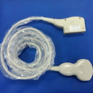
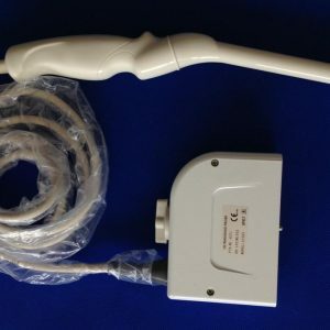
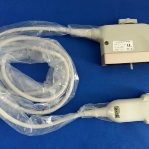
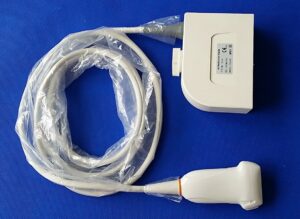
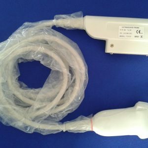
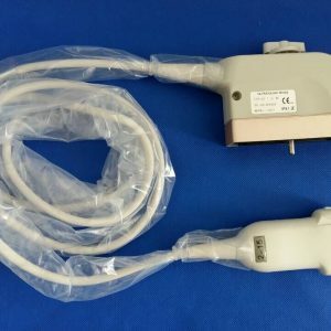
The different types of ultrasound transducers have different pros and cons, and there are many different uses for each. Read on to learn about the pros and cons of each type. You’ll also find out which type of transducer is right for you! We’ll discuss the Pros and Cons of Endocavitary and Curvilinear probes. This information will help you decide which ultrasound transducer is best for you.
Con丨Ultrasound Transducer
A con ultrasound transducer is a device used to detect soft tissue with a high resolution. The device contains a large array of crystals that are activated repeatedly to form a complete image frame. This image is called a sonogram. Because the image frame is formed 20 times a second, there is real-time motion visible on the screen. The user of the transducer must be trained to use the device safely.
To maintain the quality of the image produced by a con
ultrasound transducer, it should be properly cleaned. This means cleaning the transducer after each use, as well as using the recommended disinfecting solutions. In addition, the manufacturer should also provide guidelines on cleaning and disinfecting transducers. This will help the user maintain the image quality of the device and ensure a long product life. To disinfect the ultrasound transducer, read the instructions carefully.
An ultrasound service provider should act as a partner in the maintenance of the
ultrasound transducer. It is important to find a reliable ultrasound service provider that has the experience and expertise necessary to repair your transducer. Transducer repair is complex, but it comes down to trust and quality. Many ultrasound service providers will tout their certifications. Trisonics, for example, has achieved several certifications and is working toward higher levels. Certifications are just the beginning, however. Once the transducer has been repaired, it is important to work with the ultrasound service provider to verify the issue or to step in if it is systemic.
Pros丨Ultrasound Transducer
The main feature of an
ultrasound transducer is its ability to produce images of the inside of a human body. Ultrasound technology has made the probe more flexible than its predecessors. The ultrasound probe is adjustable, allowing it to be positioned just superior to the iliac crest, anterolaterally to the abdominal organs, and laterally to follow a structure. It can also tilt along its short axis, which allows physicians to take multiple cross-sectional images of a certain organ or structure.
The main downside of an ultrasound transducer is that it has a huge footprint, which limits the range of applications. However, if used properly, the device can produce high-quality images with excellent depth perception. However, if you’re using it to diagnose a patient, you should always check that it’s compatible with your ultrasound machine. For example, if the ultrasound transducer is not compatible with your monitor, you should try a different brand.
Another major drawback is the cost. A good ultrasound system should have an inexpensive transducer, and it’s also crucial to get a professional who has done this type of work for many years. Experienced sonographers know exactly how to manipulate the transducer and get the best results. It’s also important to choose a transducer that will not damage or kink the cable. If you drop the device, you can scratch its surface.
A recent survey of healthcare facilities revealed that the use of endocavitary ultrasound probes may pose a health risk. The study aims to assess the antimicrobial efficacy of various disinfection methods, including using a disinfectant-impregnated towel before cleaning with ultraviolet C light. These results suggest that proper disinfection practices are essential for preventing the spread of infections.
While endocavitary transducers are rarely used in the ED, their use may be justified by specific studies. Their wand-like design facilitates the examination of pelvic organs, especially the female urinary tract and the prostate. While the transabdominal examination may reveal pathology of the pelvic organs, the internal evaluation of these areas may be beneficial in diagnosing and treating certain disease states. Moreover, it can guide the drainage of peritonsilar abscesses.
This type of ultrasound probe has a curvilinear footprint and a high-frequency beam. It is important to keep the probe close to the structure to be examined. Despite its limited penetration and resolution, endocavitary
ultrasound probes are an effective option for diagnostic purposes. These
ultrasound probes are available for use in both traditional cart-based systems and handheld ultrasound devices. If you are using ultrasound on the abdomen, you should check the endocavitary probe’s durability and safety before using it.
Curvilinear probe丨Ultrasound Transducer
There are many differences between the Curvilinear probe and the Phased Array transducer. While the former is great for all-around imaging, the latter is ideal for in-depth examinations of large organs or fast-moving structures. The two types of transducers use different frequencies. Read on to learn more about the benefits of each type. The following information is based on the general differences between the two types of probes.
The curvilinear probe has been validated for use in patients with chronic respiratory disease. The curvilinear probe is more accurate than the linear whole images. The latter has less variability, but the curvilinear probe is still the preferred choice. Many ER physicians and practitioners will use a curvilinear probe to visualize intrauterine pregnancy. While it has some limitations, it is an excellent option for many applications.
A second important measurement to compare when choosing an ultrasound transducer is the field of view. This refers to the size of the image displayed by the probe when scanning. Linear probes scan straight down, while a curved array transducer uses a curved array. In most cases, a linear probe’s field of view is described in mm, whereas a curved array’s field of view is measured in degrees. Think of it as the size of a protractor.
Operating frequency丨Ultrasound Transducer
The operating frequency of ultrasound transducers is a major consideration in medical imaging. Operating frequencies range from 0.5 MHz to 100 MHz and are necessary for specific medical applications. For example, lower frequencies are used for pervasive imaging and high frequencies are used for small, close-to-the-body objects. A physician must choose the best frequency for the imaging application based on a patient’s specific needs.
A conventional ultrasound transducer’s operating frequency is typically in the range of 1 to 10 MHz. The lower frequency, as well as the f-number, is insufficient for imaging targets with high contrast in elastic properties, such as bone. Hence, ultrasound transducers with higher frequencies are preferred. The higher the frequency, the higher the resolution of the images. While a high-frequency ultrasound transducer produces images with more detail, the low-frequency model is used for imaging small structures.
Ultrasound frequencies determine image resolution and the depth of field visualized. The frequency of ultrasound transducers is inversely proportional to the depth of penetration of the ultrasound signal. A common frequency range is from 3.5 to 7.5 MHz for pelvic imaging. For transabdominal ultrasound, frequencies of 3.5 to 5 MHz are used. The same applies to transvaginal ultrasound transducers. When selecting the operating frequency, start at the lowest frequency.
Crystals丨Ultrasound Transducer
Single crystals on an ultrasound transducer are a promising candidate for high-performance ultrasound applications. These crystals exhibit a high piezoelectric and electro-mechanical coupling constant (k33>90%), making them attractive for high-resolution imaging. However, the other performance characteristics of single crystals are lower than those of PZT, including sound velocity and coercive field. This paper reviews the strengths and limitations of single crystals in medical ultrasound applications.
Piezoelectric crystals work by converting electrical pulses into mechanical vibrations. The returned mechanical vibrations can then be converted back into electrical energy. A piezoelectric crystal is a piece of material with electrodes attached on two opposite faces. This causes the crystal to expand and contract, producing mechanical motion. The motion causes the crystal plate’s thickness to expand, generating a compressional (P)-wave.
A typical crystal’s electrical impedance varies with its sensitivity, bandwidth, and impedance. This characteristic can be expressed mathematically, and it depends on the frequency and mounting configuration of the transducer. The maximum power output of a single crystal depends on the frequency and amplitude of its vibration. The maximum output is dependent on the type of crystal and its dielectric properties. Several crystals have different sensitivity, power, and bandwidth.
Image orientation丨Ultrasound Transducer
There are several ways to manipulate the
ultrasound transducer. One option is tilting, also known as fanning or sweeping. This motion holds the ultrasound transducer centered on the surface of the body. Tilting is often used to obtain serial cross-sectional images of solid organs. The other option is rocking, which involves “rocking” the transducer away or towards the probe indicator along its long axis. This motion maintains the image in plane.
The ultrasound image orientation is determined by the settings on the
ultrasound device, transducer type, and image to probe transform. If you have a transducer that is calibrated for a single image orientation, the calibration is valid for that image orientation. In general, however, the ultrasound image orientation will correspond to the transducer’s x and y axes. The ultrasound software will show a blue “U” symbol in the near corner of the image if it was calibrated for that specific orientation.
An ultrasound image of the leg reflects the aponeurosis and the attachments to the muscle fascicle. This is crucial for accurate measurement of pennation. If the image is not perpendicular to the aponeurosis, the muscle will be characterized by continuous striations. To correct this problem, the ultrasound transducer must be oriented in such a way that the distal end of the muscle is aligned with the aponeurosis.
